
A pilar cyst is a smooth, firm lump that grows under the skin of the scalp, filled with keratin from the lining of a hair follicle. They are extremely common, almost always harmless, and they tend to run in families. What they do not do is disappear on their own, and because the scalp is exactly where a comb, a hairbrush, or a pillow makes contact, even a small one can become a daily nuisance. This guide covers what causes pilar cysts, how they differ from other lumps, and everything involved in pilar cyst removal, from the procedure itself to hair regrowth and scarring.
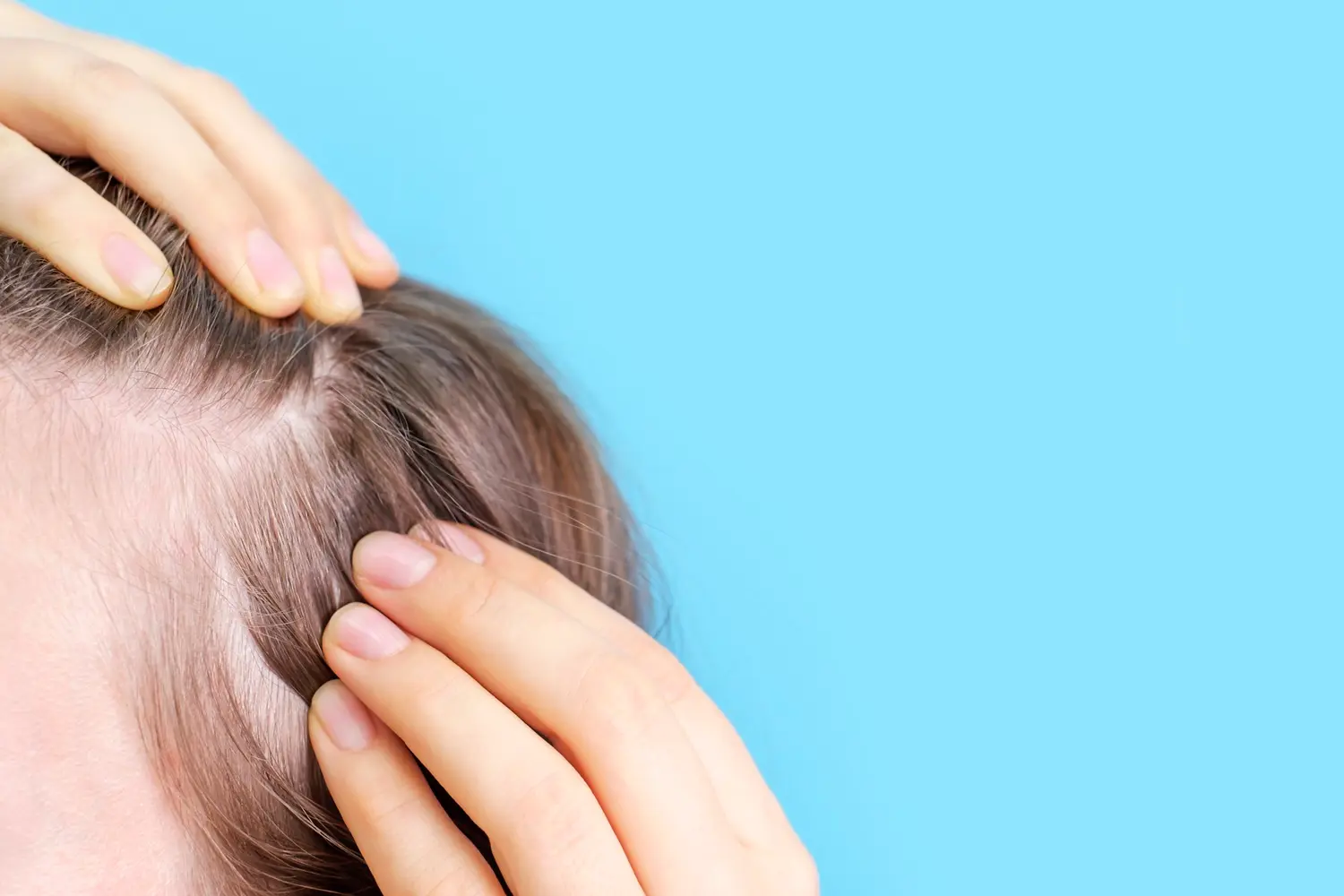
What is a pilar cyst?
A pilar cyst is a benign lump that develops beneath the skin when keratin becomes enclosed within a sac derived from a hair follicle. Keratin is the tough structural protein that makes up hair and the outer layer of skin. In a pilar cyst it accumulates inside a closed capsule, and as more is produced the lump slowly enlarges.
You may also see pilar cysts called trichilemmal cysts, isthmus-catagen cysts, or simply wens. All of these describe the same thing. The name trichilemmal refers to the trichilemma, the outer root sheath of the hair follicle, which is the specific structure the cyst wall originates from.
Because they arise from hair follicles, pilar cysts appear where follicle density is highest. Roughly 90% develop on the scalp, although they occasionally appear on the face, neck, back, or other hair-bearing areas. They are the second most common type of skin cyst after the epidermoid cyst, and unlike many lumps they are frequently multiple, with most people who get one developing several over time.
The important point is that a pilar cyst is benign. It is not a tumour in any meaningful sense, it is not contagious, and the overwhelming majority never cause anything more than inconvenience. What makes them worth treating is their location and their persistence.
Pilar cyst at a glance
- Also known as
- Trichilemmal cyst, isthmus-catagen cyst, wen.
- Usual site
- The scalp in around 90% of cases, reflecting the density of hair follicles there.
- Appearance
- Smooth, firm, dome-shaped, skin-coloured, and mobile beneath the skin. Typically no visible central pore.
- Contents
- Dense, compacted keratin, usually pale and relatively odourless compared with an epidermoid cyst.
- Who gets them
- More common in women and in middle age. They frequently run in families and are often inherited in an autosomal dominant pattern.
- Number
- Multiple in the majority of cases, so finding a second or third is common rather than unusual.
- Behaviour
- Slow-growing and benign. They do not resolve spontaneously, and around a quarter develop calcification over time.
- Treatment
- Surgical excision of the cyst together with its capsule, carried out under local anaesthetic.
Pilar cyst or epidermoid cyst?
These two are the most commonly confused skin cysts, and although both are benign and keratin-filled, they arise from different parts of the hair follicle and behave differently during removal. Telling them apart is largely a matter of site and surface.
- Around 90% occur on the scalp
- No central punctum or visible pore
- Arises from the outer root sheath of the follicle
- Contains dense, compacted, relatively odourless keratin
- Tough capsule that usually shells out intact
- Often multiple and frequently inherited
- More common on the face, neck, chest, and back
- Usually has a visible central punctum
- Arises from the follicular infundibulum
- Contains softer, cheese-like keratin with a strong odour
- More fragile wall that ruptures more readily
- Usually solitary, and less strongly hereditary
The distinction matters practically as well as academically. A pilar cyst capsule is comparatively robust, which is why an experienced clinician can often remove one whole through a surprisingly small incision. That in turn keeps the scar short and the recurrence rate low.
What causes a pilar cyst?
Pilar cysts form when cells from the outer root sheath of a hair follicle become enclosed and continue producing keratin with nowhere to shed it. Unlike many skin problems there is no lifestyle trigger to correct, and no evidence that hygiene, diet, hair products, or washing frequency play any part.
The single strongest factor. Pilar cysts often run in families and are commonly inherited in an autosomal dominant pattern, which means one affected parent can pass on the tendency.
The scalp carries far more follicles than anywhere else on the body, which is simply why the overwhelming majority of pilar cysts appear there.
They are most often diagnosed in middle age and are noticeably more common in women, although anyone can develop them.
Injury or inflammation affecting a follicle may contribute in some cases, although most pilar cysts arise with no identifiable local cause at all.
It is worth stating plainly what does not cause them, because these worries come up constantly. Pilar cysts are not caused by poor hair hygiene, by not washing your hair often enough, by shampoo or styling products, by wearing hats, by dandruff, or by anything infectious. They are not a sign that anything is wrong with your general health.
Symptoms and how to recognise one
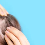
Most people discover a pilar cyst by accident, usually while washing or brushing their hair, or when a comb repeatedly catches on the same spot. The typical presentation is quite consistent.
- Smooth, round, dome-shaped lump under the skin
- Firm and rubbery rather than soft
- Mobile, moving slightly when pushed
- Skin-coloured, with normal skin over the top
- No central pore or opening
- Painless unless inflamed
- Grows very slowly, often over years
- May range from a few millimetres to several centimetres
- Frequently joined by others elsewhere on the scalp
- Hair over a large cyst may thin as the skin stretches
- Can catch on combs, brushes, and hair clippers
- May become red, tender, and swollen if it ruptures
Discomfort is not usually from the cyst itself but from what happens around it. Pressure when lying on a pillow, catching during brushing, or difficulty at the barber are the complaints that most often prompt people to seek pilar cyst removal.
Are pilar cysts dangerous?
In the overwhelming majority of cases, no. Pilar cysts are benign, they grow slowly, and they do not spread. Many people live with one for years without any consequence beyond the occasional catch of a hairbrush.
There is one uncommon variant worth knowing about. A proliferating trichilemmal tumour is a rare form in which the cyst grows unusually large, sometimes becoming lobulated, ulcerated, or rapidly enlarging. These are still usually benign, but they behave more aggressively locally and malignant change, while genuinely rare, has been described. This is the clinical reason why a scalp lump that changes character deserves proper assessment rather than continued observation.
Other scalp lumps it could be
Several other conditions can present as a lump on the scalp. Most are equally harmless, but they are managed differently, which is why an accurate diagnosis is the first step.
Very similar in feel but usually carries a visible central punctum and is more often found on the face, neck, or trunk than the scalp.
A benign fatty lump. Typically softer, more doughy, less sharply defined, and sitting deeper than a pilar cyst.
A small area of bone formation within the skin. Feels distinctly hard and immobile, quite unlike the rubbery give of a cyst.
Present from birth and typically found along lines where the facial and skull structures fused during development.
A benign growth of sweat gland origin, appearing on the scalp as a smooth pink or reddish nodule. Sometimes multiple and inherited.
Painful, hot, red, and often rapidly swollen. Requires prompt treatment of the infection before any definitive removal.
Do pilar cysts go away on their own?
They do not. This is the most important practical difference between a pilar cyst and many other skin lumps. The cyst is enclosed by a capsule that continues producing keratin, so the natural course is slow enlargement rather than resolution. Waiting does not help, and there is no cream, tablet, or home remedy that will dissolve one.
Occasionally a cyst ruptures under the skin. This can flatten the lump briefly and give the impression that it has resolved, but the capsule remains behind and the cyst almost always reforms. Rupture also causes inflammation, since keratin released into the surrounding tissue provokes a brisk reaction, so the area often becomes red, swollen, and tender for a period afterwards.
That said, treatment is not compulsory. A small, stable, symptomless pilar cyst can reasonably be left alone and monitored. Removal becomes the sensible option when it grows, gets in the way, becomes inflamed, or bothers you cosmetically.
How a pilar cyst is diagnosed
Diagnosis is usually straightforward and made clinically, from the appearance and feel of the lump together with its history. Scans are rarely necessary for a typical scalp cyst.
How long the lump has been present, how quickly it has changed, whether it has ever been inflamed, and whether other family members have had similar lumps.
Assessing whether the lump is firm, mobile, and dome-shaped, and checking for a central punctum, which points more towards an epidermoid cyst.
Examining the rest of the scalp, since pilar cysts are multiple in most cases and several can often be dealt with in one appointment.
Removed tissue is routinely sent for analysis. This confirms the diagnosis and identifies the uncommon proliferating variant if present.
Pilar cyst removal: your treatment options
Surgical removal is the only reliable treatment, and the aim in every technique is the same: to take out the entire capsule. Leaving any part of the cyst wall behind is what allows a cyst to return, so complete removal matters far more than the size of the incision.
Surgical excision
- How it worksAn incision is made over the cyst and the sac is dissected free and lifted out whole under local anaesthetic.
- Best forMost pilar cysts, including larger ones and any that have been inflamed before.
- RecurrenceLow, because the capsule is removed intact.
Minimal excision
- How it worksA small incision or punch is used, the contents are expressed, and the capsule is then teased out through the opening.
- Best forSmaller, uncomplicated cysts where a shorter scar is the priority.
- AdvantageLess scarring, provided the whole capsule is retrieved.
Drainage then excision
- How it worksAn acutely infected or inflamed cyst is drained first, with formal excision carried out later once the area has settled.
- Best forCysts that are currently infected, where immediate excision would be difficult and more likely to scar.
Because pilar cysts have a relatively tough wall, they often enucleate cleanly, meaning the whole sac lifts out in one piece. This is one reason removal from the scalp is usually quicker and tidier than people expect.
What happens during pilar cyst removal
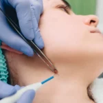
Removal is a minor procedure carried out under local anaesthetic in a single appointment, typically taking between twenty and forty minutes depending on size and number.
Preparation
The procedure
Immediately afterwards
One question comes up more than any other, so it is worth answering directly: your head will not be shaved. Only the hair immediately over the cyst needs to be parted or, at most, a very small area trimmed. Surrounding hair covers the site almost immediately, and most people find the wound is not visible to others even in the first few days.
Recovery and aftercare
Recovery is generally quick and uncomplicated. Expect some tenderness and mild swelling for two to three days, with the wound closed and comfortable within a week and sutures out at around seven to ten days. Most people return to work the same or the following day.
Do
- Keep the wound clean and dry for the first 24 to 48 hours
- Follow the advice you are given about when to wash your hair
- Take simple pain relief such as paracetamol if you need it
- Sleep on the opposite side if the site feels tender
- Attend for suture removal and ask for your histology result
- Let any scab separate naturally
Avoid
- Scratching, picking, or rubbing the wound
- Vigorous brushing or combing over the site
- Hair dye, bleach, or chemical treatments until fully healed
- Swimming and saunas until the wound has closed
- Strenuous exercise for the first couple of days
- Tight hats or headbands that press on the site
Be guided by your clinician on hair washing. Many people are advised to keep the area dry for the first day or two, then wash gently with lukewarm water and a mild shampoo, patting rather than rubbing the site dry.
Scarring and hair regrowth
Scarring after pilar cyst removal is usually minimal, and the scalp has two significant advantages. It has an excellent blood supply, which supports fast healing, and it has hair, which conceals the result. A well-placed scar within the hairline is generally undetectable once healing is complete.
Hair that was trimmed grows back normally, because the follicles themselves are not removed. A narrow line of scar tissue will not grow hair, but on a small excision this is typically too fine to notice once surrounding hair lies over it. Larger cysts require a longer incision and therefore leave a longer scar, which is one practical argument for dealing with a cyst while it is still small.
To give a scar the best chance, protect it from sun exposure while it matures, avoid tension on the area, and follow any scar care advice you are given. Scars continue to soften and fade for many months after the wound has closed.
Why pilar cysts come back
Recurrence after complete excision is uncommon, and when a cyst does return there is usually a clear explanation.
By far the most common reason. If any fragment of the cyst wall remains, it can continue producing keratin and the cyst gradually reforms.
A cyst that bursts during surgery is harder to remove completely, which is one reason previously inflamed cysts have a slightly higher recurrence rate.
Scarring from an earlier rupture or infection tethers the capsule to surrounding tissue, making clean dissection more difficult.
Often what seems to be a recurrence is simply another cyst nearby. Since pilar cysts are usually multiple, new ones developing elsewhere is expected rather than a sign that treatment failed.
Infected and inflamed pilar cysts
A pilar cyst can become inflamed, most often after the capsule ruptures beneath the skin and keratin leaks into the surrounding tissue. This provokes a strong inflammatory reaction that can look and feel very much like infection, with redness, swelling, warmth, and tenderness developing over a day or two.
Genuine bacterial infection also occurs, particularly if a cyst has been squeezed or picked at. In either case, the immediate priority is settling the inflammation rather than proceeding straight to excision, since operating on acutely inflamed tissue is more difficult, more uncomfortable, and more likely to leave a poor scar.
Treatment may involve antibiotics where infection is present, or drainage to relieve pressure if the area is tense and painful. Definitive removal is then planned for several weeks later, once the tissue has settled and the capsule can be dissected cleanly.
Risks and possible complications
Pilar cyst removal is a low-risk procedure, but as with any minor surgery it is sensible to know what is normal and what is not.
- Tenderness and mild swelling for a few days
- Slight bruising around the site
- A small amount of bleeding on the first day
- A firm ridge under the scar that softens over weeks
- Wound infection
- Recurrence if capsule fragments remain
- A more noticeable or widened scar
- Temporary altered sensation around the site
When to seek professional advice
FAQs about pilar cysts
Are pilar cysts cancerous?
No. Pilar cysts are benign. A rare variant called a proliferating trichilemmal tumour can grow large and behave more aggressively locally, and malignant change has been described but is genuinely rare. Removed tissue is routinely sent for analysis to confirm the diagnosis.
Will a pilar cyst go away on its own?
No. The capsule keeps producing keratin, so the natural course is slow growth rather than resolution. A cyst that ruptures may flatten temporarily, but it almost always reforms because the capsule remains.
Can I squeeze or pop a pilar cyst?
No. Squeezing can rupture the capsule under the skin, causing inflammation, pain, and possible infection. It leaves the capsule in place so the cyst returns, and the resulting scar tissue makes proper removal harder later.
Will my head be shaved for the procedure?
No. The hair is usually just parted, and at most a very small area immediately over the cyst is trimmed. Surrounding hair covers the site almost straight away.
Does pilar cyst removal hurt?
The procedure itself should not hurt, since the area is fully numbed with local anaesthetic. You will feel pressure and movement. Afterwards most people describe mild soreness that responds to simple pain relief.
How long does recovery take?
Tenderness and mild swelling usually last two to three days. Sutures are typically removed after seven to ten days, and most people return to work the same or the next day.
When can I wash my hair afterwards?
Most people are advised to keep the area dry for the first day or two, then wash gently with lukewarm water and a mild shampoo. Follow the specific advice your clinician gives you.
Will it leave a scar?
There will be a small scar, but on the scalp it is usually concealed by hair and often undetectable once healed. Smaller cysts need shorter incisions, which is a good reason not to leave one to grow.
Why do I keep getting pilar cysts?
Because they are usually multiple and frequently inherited. Developing several over the years is typical and does not mean anything has gone wrong with previous treatment.
Can pilar cysts appear anywhere other than the scalp?
Yes, though it is much less common. Around 90% occur on the scalp, with the remainder appearing on the face, neck, back, and other hair-bearing areas.
Is removal available on the NHS?
Removal is often considered a cosmetic procedure unless the cyst is symptomatic, infected, or rapidly changing, so availability varies. Many people choose private treatment for quicker access and greater control over timing.
Conclusion
Pilar cysts are common, benign, and strongly hereditary, but they share one stubborn characteristic: they do not go away by themselves. A small, settled cyst can safely be left alone and monitored. Anything that is growing, catching on a brush, becoming inflamed, or simply bothering you is straightforward to deal with, and dealing with it sooner generally means a smaller incision and a finer scar.
The single factor that determines success is complete removal of the capsule. That is what prevents the cyst from returning, and it is why squeezing or draining a cyst at home achieves nothing lasting while making later surgery more difficult.
At Linia Skin Clinic, cyst removals are carried out by our experienced dermatology team with a focus on minimal downtime and the best possible cosmetic result. Our Medical Director, Dr Simon Zokaie, qualified from the University of Manchester in 2003, holds MRCP membership, and specialises in cosmetic dermatology with an emphasis on minimal-downtime procedures.
If you have a lump on your scalp you would like assessed or removed, book a consultation with our team and we will recommend the most appropriate approach for you.