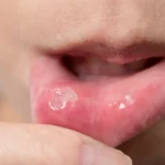

An oral mucocele is a soft, painless, bluish or translucent swelling that appears inside the mouth, most often on the inner surface of the lower lip. It forms when a minor salivary gland is damaged or blocked and mucus collects in the surrounding tissue. Mucoceles are harmless and some settle by themselves within a few weeks, but those that persist, recur, or keep getting in the way of eating and speaking are usually best dealt with by oral cyst removal. This guide explains what causes an oral mucocele, how it is diagnosed, every removal option available, and what recovery genuinely involves.
What is an oral mucocele?
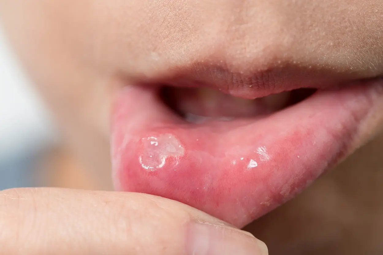
An oral mucocele, also called a mucous cyst or mucocoele, is a swelling that develops when mucus escapes from, or becomes trapped within, one of the hundreds of minor salivary glands lining the mouth. These tiny glands sit just beneath the surface of the lips, cheeks, tongue, and palate, and each drains through a very fine duct. If that duct is severed or obstructed, saliva has nowhere to go and pools in the surrounding tissue, producing a soft, dome-shaped lump.
Most oral mucoceles are between 1mm and 10mm across, although they can grow larger. They typically look bluish and translucent when they sit close to the surface, and more skin-coloured or pale when they lie deeper in the tissue. They are soft and fluctuant to the touch, meaning they feel as though they contain fluid, because they do.
Crucially, a mucocele is benign. It is not a tumour, it is not contagious, and it does not turn into anything sinister. What makes it worth addressing is not danger but nuisance: a lump on the inner lip sits exactly where the teeth meet, so it is repeatedly bitten, which keeps the cycle going.
Strictly speaking, the most common form is not a true cyst at all. Because it has no epithelial lining (the mucus simply spills into the tissue and the body walls it off with granulation tissue), pathologists classify it as a pseudocyst. This distinction matters more than it sounds, because it explains why draining one rarely fixes it.
Types of oral mucocele
Mucoceles are grouped by the mechanism that created them and by where they form. The type influences how straightforward oral cyst removal will be, and how likely the lesion is to return.
Mucous extravasation cyst. By far the most common type, accounting for the majority of cases. The duct of a minor salivary gland is torn, almost always through trauma such as lip biting, and mucus leaks into the surrounding connective tissue. It has no epithelial lining. Most often found on the lower lip, and most common in children and young adults.
Mucous retention cyst. Less common. Here the duct is not torn but blocked, often by a small salivary stone or by scarring, so saliva backs up and dilates the duct itself. This type does have an epithelial lining, making it a true cyst. It tends to occur in older adults and favours the floor of the mouth, the palate, and the buccal mucosa.
Ranula. A mucocele arising from the sublingual gland in the floor of the mouth. Ranulas are usually larger than lip mucoceles and have a characteristic translucent, bluish appearance often likened to a frog’s belly, which is where the name comes from. They can interfere with tongue movement, speech, and swallowing.
Plunging (cervical) ranula. An uncommon variant in which the mucus tracks downwards through or around the mylohyoid muscle and presents as a swelling in the neck, sometimes with little or nothing visible inside the mouth. This type needs specialist assessment and imaging, and is managed differently from a simple lip mucocele.
Superficial mucocele. Small, fragile blisters sitting immediately beneath the surface, usually on the soft palate, retromolar area, or inside the cheek. They rupture easily, often go unnoticed, and are more likely to appear in crops. They are sometimes associated with inflammatory conditions such as lichen planus.
What causes an oral mucocele?
The underlying cause is nearly always mechanical damage to a minor salivary gland duct. Many people can pinpoint the moment it happened, such as a catch of the teeth on the lip while eating or talking, but just as many cannot, because the initial trauma is minor and the swelling appears days later.
The single most common trigger. An accidental bite, or a habit of chewing the inner lip when concentrating or anxious, severs the delicate duct beneath the surface.
Repetitive suction on the inner lip, common in children and in people under stress, applies enough shearing force to damage a duct over time.
Sharp or chipped teeth, ill-fitting dentures, orthodontic brackets, and dental appliances can rub against the mucosa and injure the gland repeatedly.
Lip and tongue piercings sit directly over minor salivary glands and are a well-recognised cause of both initial mucoceles and stubborn recurrences.
In retention-type mucoceles a small salivary stone (sialolith) or scar tissue blocks the duct, so saliva backs up rather than leaking out.
Any procedure or laceration in the area can leave scarring that distorts a duct, which is one reason a mucocele sometimes returns close to the site of an earlier removal.
It is worth saying plainly what does not cause a mucocele: it is not a sign of poor oral hygiene, it is not an infection, it is not a sexually transmitted condition, and it is not caused by anything you have eaten. These are all common worries, and none of them holds up.
Symptoms and how to recognise one
Oral mucoceles have a fairly distinctive presentation, which is why an experienced clinician can often identify one on sight. The classic picture looks like this:
- Soft, smooth, dome-shaped swelling
- Bluish, translucent, or pearly grey when superficial
- Painless in the vast majority of cases
- Fluctuant, giving slightly under gentle pressure
- Usually 1mm to 10mm across
- Appears over a few days rather than instantly
- May grow and shrink, sometimes over weeks
- Can rupture, release clear or straw-coloured fluid, then refill
- Often becomes firmer and paler if it has been present for months
- May be repeatedly traumatised by the teeth
- Can interfere with chewing, speech, or wearing dentures
Pain is not a typical feature. If a lump in the mouth is genuinely painful, rapidly enlarging, bleeding, ulcerated, firm and fixed to deeper tissue, or accompanied by numbness, it warrants prompt assessment. Those features point away from a simple mucocele.
Where mucoceles appear in the mouth
Location is genuinely diagnostic. Minor salivary glands are distributed throughout the mouth, but mucoceles cluster in predictable places, and one location in particular changes the clinical thinking entirely.
- Lower lip
- The commonest site by a wide margin, accounting for most reported cases. The inner surface of the lower lip sits directly in the path of the upper teeth, so it takes the brunt of accidental biting.
- Floor of the mouth
- Mucoceles here arise from the sublingual gland and are termed ranulas. They are typically larger, more translucent, and more likely to affect tongue movement and swallowing.
- Ventral tongue
- The undersurface of the tongue contains the glands of Blandin-Nuhn. Mucoceles here are less common, move with the tongue, and are easily irritated by the lower teeth.
- Inner cheek
- Buccal mucosa mucoceles usually follow cheek biting or friction from a sharp tooth or dental appliance, and often sit along the line where the teeth meet.
- Soft palate and retromolar area
- Typically superficial mucoceles: small, fragile, and prone to rupturing before they are ever examined.
- Upper lip
- Uncommon, and treated with more caution. A persistent swelling in the upper lip is statistically more likely to be a minor salivary gland tumour than a mucocele, so lesions here are generally excised and sent for histological examination rather than simply observed.
Other oral lumps it could be
Several other lesions can look similar to an oral mucocele, particularly once a mucocele has been present for a while and has become firmer. This is precisely why a lump that does not settle should be assessed rather than assumed.
A firm, pale, fibrous nodule caused by chronic irritation. Similar location and history, but solid rather than fluid-filled and does not fluctuate in size.
A soft, yellowish swelling of fatty tissue. Usually deeper, slower-growing, and lacking the bluish translucency of a mucocele.
A vascular lesion that blanches, or goes pale, when pressed with a glass slide. A mucocele does not blanch, which is a useful bedside distinction.
Both benign and malignant salivary tumours can present as a painless lump, especially on the upper lip and palate. This is the reason removed tissue is routinely sent for analysis.
Painful, warm, red, and often accompanied by a bad taste or systemic symptoms, which is a very different clinical picture requiring different treatment.
A small, firm, yellowish-white nodule typically found in the floor of the mouth or on the tonsillar pillar. Harmless, but easily confused with a mucocele.
Will an oral mucocele go away on its own?
Sometimes, yes. A proportion of small, recently formed mucoceles, particularly superficial ones on the lip in children and young adults, rupture spontaneously and resolve within roughly two to eight weeks as the damaged duct heals and the trapped mucus is reabsorbed. If a lump is small, painless, and only appeared a week or two ago, a period of watchful waiting is entirely reasonable.
What tends not to resolve is the mucocele that has been present for months, that has ruptured and refilled several times, or that sits where the teeth catch it repeatedly. Each cycle of rupture and refill lays down more fibrous tissue, so the lesion gradually becomes firmer, paler, and less likely to disappear. Ranulas in the floor of the mouth rarely resolve without treatment.
The reason for the difference is straightforward. If the mucus drains but the damaged gland continues to produce saliva into the same space, the swelling simply returns. Definitive treatment addresses the gland, not just the fluid, which is exactly the principle behind surgical oral cyst removal.
How an oral mucocele is diagnosed
In most cases diagnosis is clinical, made from the appearance, the location, and the history of the lump, without any need for scans or blood tests. A typical assessment involves:
When the lump appeared, whether it has changed in size, whether it has ever burst, and whether you recall biting or injuring the area.
Inspecting the colour and surface, palpating to establish whether it is soft and fluctuant or firm and fixed, and checking whether it blanches under pressure.
Looking for the source of the trauma, such as a sharp tooth edge, a piercing, or an ill-fitting denture, because leaving that in place invites recurrence.
Rarely needed for lip mucoceles. Ultrasound or MRI is used for larger ranulas, suspected plunging ranulas, or where the extent of a lesion is unclear.
When a mucocele is removed, the tissue is normally sent for histopathological examination. This is routine rather than a cause for concern: it confirms the diagnosis definitively and rules out the small number of lesions, particularly on the upper lip and palate, that can mimic a mucocele.
Oral cyst removal: your treatment options
Several approaches to oral cyst removal exist, and the right one depends on the size of the lesion, where it sits, how many times it has recurred, and the age of the patient. What they all share is the aim of removing or destroying the offending salivary gland tissue rather than simply emptying the swelling.
Excision with gland removal
- How it worksUnder local anaesthetic, the lesion is removed together with the associated minor salivary gland and any adjacent glands that feed it.
- Best forPersistent or recurrent lip, cheek, and tongue mucoceles. The established standard.
- RecurrenceLowest of all methods, because the source gland is taken out.
Laser ablation
- How it worksA CO2 or diode laser vaporises the lesion while sealing small vessels as it goes.
- Best forSmall, well-defined mucoceles, and for patients where minimal bleeding is a priority.
- AdvantagesLittle bleeding, often no sutures, and generally comfortable healing.
Marsupialisation
- How it worksThe lesion is opened and its edges stitched to the surrounding mucosa so it drains continuously and shrinks.
- Best forLarger ranulas in the floor of the mouth, where full excision risks nearby structures.
- Trade-offLess invasive, but a higher recurrence rate than excision.
Micro-marsupialisation
- How it worksA suture is passed through the widest part of the lesion and left in place for about a week, creating a drainage tract.
- Best forChildren and anxious patients. Quick, minimally invasive, and well tolerated.
Cryosurgery
- How it worksThe lesion is frozen, destroying the abnormal tissue, which then sloughs away as the area heals.
- Best forSmall superficial mucoceles where surgery is impractical.
Intralesional treatment
- How it worksCorticosteroid or a sclerosing agent is injected to shrink the lesion and encourage the cavity to close.
- Best forSelected cases, including some ranulas, or where surgery is best avoided.
For a straightforward mucocele on the lower lip, excision under local anaesthetic remains the most predictable option, and is typically completed in a single appointment lasting around twenty to thirty minutes.
What happens during oral cyst removal
Knowing the sequence in advance makes the appointment considerably less daunting. A typical excision of a lip mucocele runs as follows:
Preparation
The procedure
Immediately afterwards
You should feel pressure and movement but no pain during the procedure. Most people drive themselves home afterwards, since local anaesthetic rather than sedation is used, and return to work the same or the following day.
Recovery and aftercare
Recovery from oral cyst removal is generally quick. The mouth heals faster than skin elsewhere on the body thanks to its rich blood supply, and most people find discomfort is mild and short-lived. Expect some swelling and tightness in the lip for two to three days, with the site settling over one to two weeks and any dissolvable sutures disappearing within roughly seven to ten days.
Do
- Rinse gently with warm salt water from the day after, especially after meals
- Stick to soft, cool foods for the first 48 hours
- Take simple pain relief such as paracetamol if you need it
- Brush your teeth as normal, working carefully around the site
- Use a cold compress on the outside of the lip for the first day
- Attend any follow-up appointment and ask for your histology result
Avoid
- Poking or stretching the area to inspect it
- Hot, spicy, acidic, crunchy, or very salty foods early on
- Smoking and alcohol, both of which slow mucosal healing
- Vigorous rinsing or spitting in the first 24 hours
- Strenuous exercise for the first day or two
- Returning to lip biting or nibbling once healed
A small firm ridge of scar tissue where the lesion was is normal and softens over several weeks. Numbness of a small patch of lip can occur if a tiny sensory nerve branch was disturbed; this is uncommon and almost always temporary.
Why mucoceles come back
Recurrence is the most common frustration with oral mucoceles, and it nearly always has an identifiable reason. Understanding these makes it far easier to have a useful conversation with your clinician:
Draining the fluid or removing only the lining leaves the damaged gland still producing saliva into the same space. This is the single biggest cause of recurrence.
Minor salivary glands sit in clusters. Surgery can occasionally injure a neighbouring duct, producing a new mucocele close to the original site.
If a sharp tooth, denture edge, or piercing caused the problem and stays unaddressed, the same injury simply happens again.
Ongoing lip biting or sucking re-traumatises the healed area. Addressing the habit is part of the treatment, not an afterthought.
Where a mucocele has recurred more than once, it is reasonable to ask whether the approach should change, for example moving from marsupialisation to formal excision, or in the case of a repeatedly recurring ranula, considering removal of the sublingual gland itself.
Risks and possible complications
Oral cyst removal is a minor, low-risk procedure, but no operation is entirely without risk and you should know what to look for.
- Swelling and tightness for a few days
- Mild soreness once the anaesthetic wears off
- A small amount of bleeding on the first day
- A temporary firm ridge at the site
- Recurrence of the mucocele
- Infection of the surgical site
- Temporary numbness of a small area of lip
- A visible or palpable scar, usually minimal inside the mouth
Mucoceles in children
Oral mucoceles are common in children and adolescents, largely because lip biting and lip sucking are so prevalent at those ages. The good news is that paediatric mucoceles have a relatively high rate of spontaneous resolution, so a short period of observation is often the first step rather than immediate surgery.
Where treatment is needed, micro-marsupialisation is frequently preferred in younger children. It involves passing a single suture through the lesion under local anaesthetic, takes only a few minutes, requires no incision, and is far better tolerated than a formal excision, while still producing good results in appropriately selected cases.
For parents, the most valuable contribution is usually addressing the underlying habit. Gently discouraging lip sucking and nibbling, and having a dentist smooth any sharp tooth edge or adjust an orthodontic appliance that is rubbing, does more to prevent recurrence than any procedure.
When to seek professional advice
FAQs about oral mucoceles
Is an oral mucocele dangerous or cancerous?
No. An oral mucocele is a benign, non-contagious swelling caused by a damaged salivary gland duct, and it does not become cancerous. Removed tissue is routinely sent for analysis simply to confirm the diagnosis, because a small number of other lesions can look similar.
Can I pop an oral mucocele at home?
No. Popping or lancing it may flatten the swelling briefly, but the damaged gland keeps producing saliva so it refills within days. Self-drainage risks infection, bleeding, and scarring, and makes later removal more difficult.
How long does an oral mucocele take to go away?
Some small, recent mucoceles resolve on their own within two to eight weeks. Those that have been present for months, have repeatedly ruptured and refilled, or sit in the floor of the mouth are unlikely to disappear without treatment.
Does oral cyst removal hurt?
The procedure itself should not hurt, as the area is fully numbed with local anaesthetic. You will feel pressure and movement. Afterwards, most people describe mild soreness that responds well to simple pain relief.
How long is recovery after oral cyst removal?
Swelling and tightness typically last two to three days, with the site settling over one to two weeks. Dissolvable sutures usually disappear within seven to ten days. Most people return to work the same or the next day.
Will the mucocele come back after removal?
Recurrence is uncommon when the lesion is excised together with the associated salivary gland. It is more likely if the fluid was simply drained, if a neighbouring duct was disturbed, or if the original cause, whether a sharp tooth, a piercing, or a lip-biting habit, has not been addressed.
Will it leave a scar?
Scarring inside the mouth is usually minimal, as oral mucosa heals particularly well. A small firm ridge at the site is normal initially and softens over several weeks.
What is the difference between a mucocele and a ranula?
They are the same process in different places. A ranula is a mucocele arising from the sublingual gland in the floor of the mouth. Ranulas tend to be larger, more translucent, more likely to affect speech and swallowing, and less likely to resolve without treatment.
Can I eat and drink normally afterwards?
Wait until the anaesthetic has fully worn off, usually two to four hours, so you do not accidentally bite the numb area. Stick to soft, cool foods for the first couple of days and avoid hot, spicy, acidic, or crunchy items while the site settles.
Are oral mucoceles caused by poor oral hygiene?
No. They are caused by mechanical damage to a minor salivary gland duct, most often from lip biting. Oral hygiene, diet, and infection are not the cause, though good hygiene does support healing after removal.
Conclusion
An oral mucocele is a benign and very common condition, but it is also a persistent one when left to its own devices. Small, recent lesions are reasonable to watch for a few weeks. Anything that has lasted longer than a month, keeps bursting and refilling, or is repeatedly caught by the teeth is unlikely to settle on its own, and the definitive answer is oral cyst removal that takes out the damaged salivary gland rather than simply emptying the swelling.
The two things that matter most are an accurate diagnosis and complete removal. The first rules out the small number of lesions that can imitate a mucocele; the second is what stops it coming back. Neither is achievable by draining a lump at home, which is why a persistent oral swelling is always worth having properly assessed.
If you have a lump inside your mouth that will not settle, arrange an assessment with a suitably qualified clinician who can confirm what it is and recommend the most appropriate removal method for its size and location.