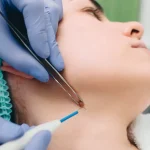

Epidermoid and pilar cysts are the two lumps most often mistaken for one another, and both are routinely called a “sebaceous cyst” even though neither comes from an oil gland. They look almost identical from the outside: a smooth, firm swelling under intact skin, filled with keratin. What separates them is where they grow, what lines them, and how likely they are to run in families. Getting the label right matters, because it changes where the lump tends to appear, how it behaves, and what to expect from removal. This guide compares the two side by side, explains how to tell them apart, and points you to the right treatment page for each.
What each cyst actually is
Both are true keratin cysts, and both are frequently mislabelled. An epidermoid cyst, also called an epidermal inclusion cyst, forms when surface skin cells become sealed inside a pocket in the dermis. That pocket is lined with the same stratified squamous epithelium as your outer skin, and it carries on producing keratin with nowhere to drain, so the cyst slowly enlarges.
“Removing the entire cystic lining is important in decreasing recurrence.”
Zito PM, Scharf R. Epidermoid Cyst. StatPearls, published by the National Library of Medicine, NIH
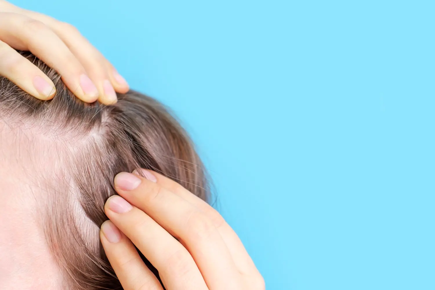
A pilar cyst, also called a trichilemmal or wen, arises instead from the outer root sheath of the hair follicle. Because that structure is densest on the scalp, this is where pilar cysts overwhelmingly appear. Their wall is thicker and tougher than an epidermoid cyst’s, which is why a pilar cyst can often be removed intact in one piece. According to StatPearls, pilar cysts are the second most common cutaneous cyst after epidermoid cysts, affect roughly 5% to 10% of the population, are more common in women, and frequently show an autosomal dominant, familial pattern.
Neither contains oil or pus. Both are filled with compacted keratin, which is why the term “sebaceous cyst” is misleading for both: a true sebaceous cyst, arising from an oil gland, is far rarer than either of these.
Epidermoid vs pilar cyst at a glance
The two overlap enough that they are routinely confused, but they differ on almost every feature that matters clinically. The table below sets them side by side.
| Feature | Epidermoid cyst | Pilar cyst |
|---|---|---|
| Origin | Surface epidermis / follicular infundibulum | Outer root sheath of the hair follicle |
| Commonest site | Face, neck and trunk | Scalp, in around 90% of cases |
| Relative frequency | Most common cutaneous cyst | Second most common |
| Who it affects | Around twice as common in men | More common in women |
| Inheritance | Usually sporadic | Often familial, autosomal dominant |
| Central punctum | Often present | Usually absent |
| Sac wall | Thinner, ruptures more easily | Thicker, often shells out intact |
| Contents | Compacted keratin, may smell | Dense keratin |
| Malignant change | Around 1% of cases | Rare (proliferating trichilemmal tumour) |
None of these single features is decisive on its own, but taken together they usually settle which cyst you are looking at. Site does most of the work: a lump on the hairy scalp is far more likely to be pilar, while the same lump on the face, neck or trunk is far more likely to be epidermoid.
How to tell them apart, and from other lumps
Beyond each other, both cysts are also confused with a handful of other lumps. Here is how the common ones behave.
Smooth, mobile, firm, under intact skin on the face, neck or trunk. Often has a tiny central punctum, the blocked pore it grew from. Painless unless inflamed.
Smooth, firm and mobile like an epidermoid cyst but almost always on the scalp, usually without a punctum, and often more than one. Frequently runs in families.
Soft, doughy, slow growing and deeper than a cyst, with no punctum. The skin glides freely over it. Covered on our lipoma page.
Tiny white keratin cysts a millimetre or two across, usually on the cheeks or around the eyes. Essentially miniature epidermoid cysts, dealt with by simple de-roofing.
Red, hot, painful and fast growing over days rather than months. This is active infection, not a quiet cyst, and it needs drainage rather than planned removal.
Present from birth and typically along developmental fusion lines such as the outer eyebrow. Firmer and more fixed, and assessed before removal because some sit deeper than they appear.
Why they form
The two cysts start from different structures, so their triggers differ too.
- Epidermoid: blocked follicles
- A hair follicle that seals over while cells continue to shed inside is the standard mechanism. This is why follicle-rich areas such as the face, neck and trunk are the commonest sites.
- Epidermoid: acne and trauma
- Inflammation from previous acne or folliculitis can leave a sealed pocket behind, and any injury that pushes surface cells into the dermis, such as a cut or piercing, can seed an implantation cyst.
- Pilar: the hair root sheath
- Pilar cysts arise from the outer root sheath rather than the surface, which is why they concentrate on the scalp where that structure is densest.
- Pilar: inheritance
- Pilar cysts frequently run in families in an autosomal dominant pattern, which is why they are often multiple and why several members of one family may have them.
- Genetic tendency, either type
- Some people simply form multiple cysts. Where cysts are numerous or start young, an inherited syndrome such as Gardner syndrome is occasionally relevant, though this is uncommon.
- Often no clear cause
- In many cases a cyst appears without any identifiable trigger. What matters is not the cause but whether it is causing problems and whether removal is appropriate.
Do they need removing?
Neither cyst has to be treated if it is not troubling you. The NHS is explicit on this point:
“Skin cysts do not need to be treated if they’re not causing any problems.”
NHS, Skin cyst
But leaving a cyst alone is not the same as it disappearing. Both epidermoid and pilar cysts have a living lining, so they persist and usually grow slowly. And leaving one alone is only reasonable once you know what it is:
“See a GP if you have a lump on your skin and you do not know what it is.”
NHS, Skin cyst
The one thing to avoid in the meantime applies equally to both:
“Do not squeeze a skin cyst. If it bursts it could become infected, or if it’s already infected you might spread the infection.”
NHS, Skin cyst
Consider removal when the cyst is growing, has become inflamed or infected more than once, catches on a comb, collar or razor, is visible enough to bother you, or the diagnosis is not certain.
How each is treated
The principle is the same for both cysts and it is simple to state: the whole sac wall has to come out, because any lining left behind can refill. Draining a cyst without removing its lining relieves pressure but does not cure it. Pilar cysts, with their thicker wall, often lift out in one piece; epidermoid cysts have a thinner wall that needs careful handling to remove intact.
Watch and manage
- ObservationReasonable for a small, stable, painless cyst with a confirmed diagnosis.
- Warm compressEases discomfort in an inflamed cyst. Does not remove it.
- Photograph monthlyThe most reliable way to track slow growth.
Definitive removal
- Complete surgical excisionWhole sac removed. Lowest recurrence for both cyst types.
- Minimal or punch excisionSmaller scar, slightly higher recurrence.
- Incision and drainageFor acute infection only. Lining stays, so it refills.
Scar care
- Silicone gel or sheetingApplied once the wound has fully closed.
- Incision along skin linesA scar hidden in a natural crease is far less visible.
- Sun protectionA new scar darkens easily in sunlight.
Risks, scarring and recurrence
- Recurrence
- For both cysts, recurrence depends on whether the lining is fully removed. Complete excision has the lowest recurrence; after simple incision and drainage, where the lining is left in place, refilling should be regarded as the expected outcome rather than a complication.
- Scarring
- Any incision can scar. On the scalp a pilar cyst scar is usually hidden by hair; on the face, neck or trunk, placing the incision along a natural skin crease does more for the final appearance than incision length alone.
- Infection
- Uncommon after planned excision of a quiet cyst. Risk rises sharply if the cyst is operated on while actively inflamed, which is why surgery is deferred until it settles.
- New cysts
- Removing one cyst does not stop a cyst-prone person forming others, and this is especially true of familial pilar cysts. That is a tendency of the skin, not a failure of the operation.
When a lump needs more than reassurance
The overwhelming majority of both cysts are entirely benign. A small minority are not, and the epidermoid figures are worth stating plainly:
“Approximately 1% of epidermoid cysts have been noted to have a malignant transformation.”
Zito PM, Scharf R. Epidermoid Cyst. StatPearls, published by the National Library of Medicine, NIH
Malignant change in a pilar cyst, into a proliferating trichilemmal tumour, is rare but recognised. In both cases the practical response is the same: removed tissue is sent for histology as a matter of routine, because the outside appearance of a cyst is a poor guide to what the lining is doing.
Both conditions, and related lumps, are covered on our skin cysts page.
FAQs
What is the difference between an epidermoid and a pilar cyst?
Both are keratin-filled cysts, but they start from different structures. An epidermoid cyst arises from the surface layer of skin and appears mostly on the face, neck and trunk. A pilar cyst arises from the hair root sheath and forms on the scalp in around 90% of cases, is more common in women, and often runs in families.
Is a pilar cyst the same as a sebaceous cyst?
No. “Sebaceous cyst” implies an oil gland, but a pilar cyst is filled with keratin, not sebum. The same applies to epidermoid cysts. Most lumps casually called sebaceous are in fact one of these two keratin cysts.
How can I tell which one I have?
Site is the strongest clue. A smooth, firm lump on the hairy scalp, usually without a central pore and sometimes one of several, is most likely a pilar cyst. The same lump on the face, neck or trunk, often with a tiny central punctum, is most likely an epidermoid cyst. A clinician can usually confirm it on examination.
Will either cyst go away on its own?
No. Both are lined with living cells that keep producing keratin, so neither has a mechanism for resolving. They may stay the same size for years, but they will not disappear. Only removal of the lining clears them permanently.
Which is easier to remove?
Pilar cysts have a thicker, tougher wall and often lift out intact in one piece. Epidermoid cysts have a thinner wall that needs careful handling to remove whole. Either way, taking out the entire sac is what prevents it coming back. See the dedicated epidermoid and pilar cyst removal guides for the detail.
Can I squeeze or pop either cyst at home?
No. The NHS advises against squeezing any skin cyst, because rupture risks infection and spreads the contents. It also leaves the sac behind so the cyst refills, and inflames the tissue, making later removal harder and recurrence more likely.
Can these cysts be cancerous?
Rarely. Around 1% of epidermoid cysts show malignant transformation, and pilar cysts can rarely become a proliferating trichilemmal tumour. This is why removed tissue is sent for histology routinely, and why a lump that grows quickly, becomes fixed or ulcerates should be assessed promptly.
Do pilar cysts run in families?
Often, yes. Pilar cysts frequently show an autosomal dominant, familial pattern, which is why they are commonly multiple and why several members of one family may have them. Epidermoid cysts are usually sporadic by comparison.
Sources
- Zito PM, Scharf R. Epidermoid Cyst. StatPearls Publishing. National Library of Medicine, National Institutes of Health.
- Al Aboud DM, Yarrarapu SNS, Patel BC. Pilar Cyst. StatPearls Publishing. National Library of Medicine, National Institutes of Health.
- NHS. Skin cyst. National Health Service, United Kingdom.
This article is for information only and does not replace an assessment in person. If you have a lump on your skin or scalp that you cannot account for, please see a clinician.