
A lump in the earlobe is almost always an epidermoid cyst, a pocket of trapped keratin rather than a growth. This guide explains what is actually inside it, why the earlobe is such a common site, how each removal method compares on recurrence and scarring, and the small number of lumps that need histology rather than simple drainage.
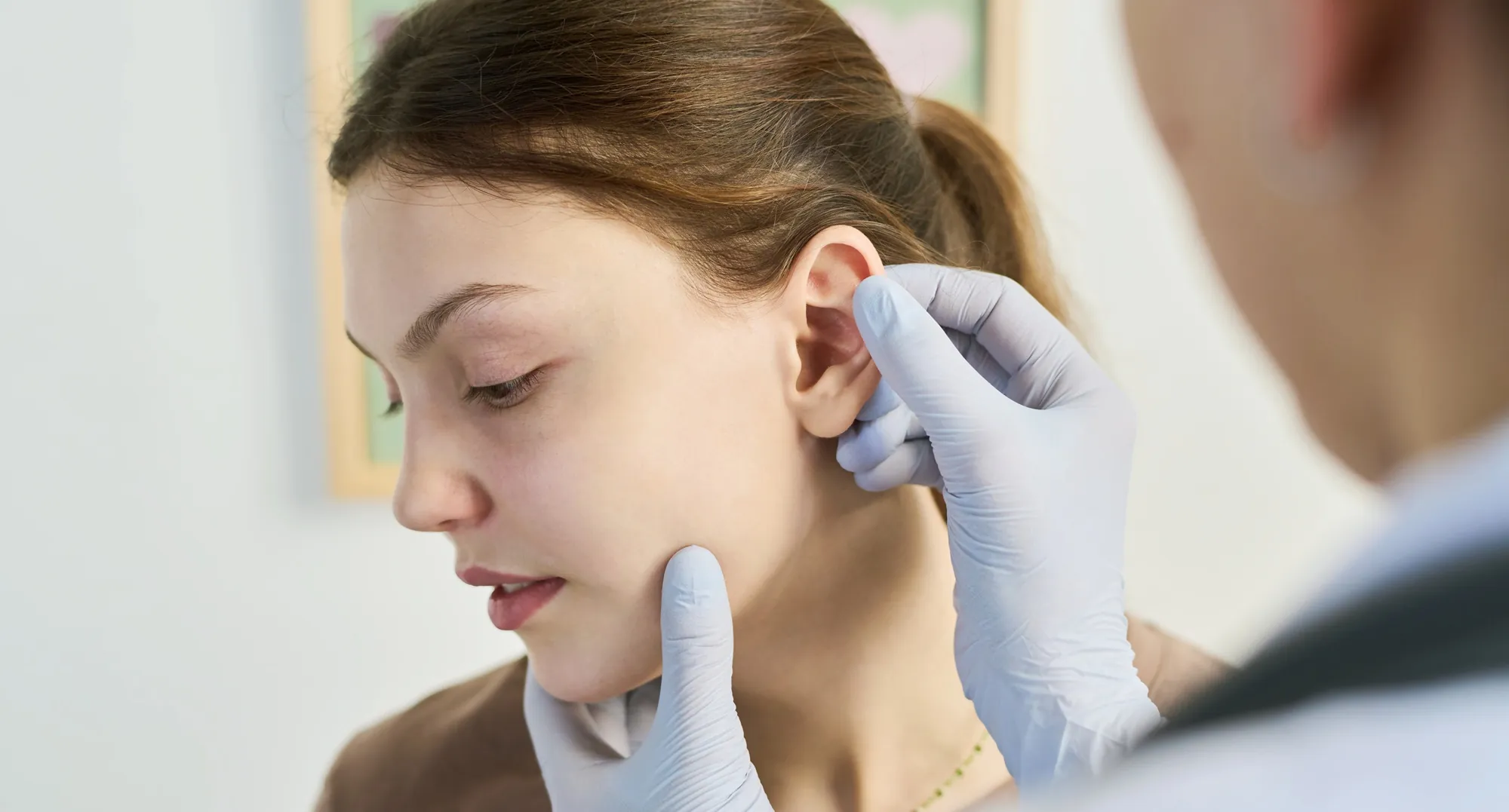
What an earlobe cyst actually is
The lump most people call a sebaceous cyst on the ear is usually an epidermoid cyst, also known as an epidermal inclusion cyst. Skin cells that should have shed from the surface become sealed inside a pocket in the dermis. That pocket is lined with the same stratified squamous epithelium as your outer skin, and it keeps doing what skin does: producing keratin. The keratin has nowhere to go, so the cyst slowly fills and enlarges.
Two consequences follow from that structure, and they explain almost everything about treatment. First, the cyst cannot resolve by itself, because the lining is still alive and still producing. Second, if you drain the contents but leave the lining behind, the pocket refills.
“Removing the entire cystic lining is important in decreasing recurrence.”
Zito PM, Scharf R. Epidermoid Cyst. StatPearls, published by the National Library of Medicine, NIH
The contents are not oil or pus. They are compacted keratin, which is why a cyst squeezed at home produces a thick, pale, strong smelling material rather than the clear fluid people expect.
The four lumps people call an earlobe cyst
They look similar from the outside and are managed very differently. Getting the label right is the whole diagnosis.
Smooth, mobile, firm, sitting under intact skin. Often has a tiny central punctum, the blocked pore it grew from. Painless unless it becomes inflamed. This is the classic earlobe cyst.
A shiny, rubbery overgrowth of scar tissue at a piercing site, extending beyond the original wound. It is scar, not a sac, and excision alone is not enough to control it.
Hot, red, tender and swelling quickly. The cyst wall has ruptured and the keratin is irritating surrounding tissue. Needs settling before any planned excision.
Soft, doughy, slow growing and deeper than a cyst, with no punctum. Less common in the earlobe than in the neck, but worth excluding on examination.
Why the earlobe in particular
Earlobes attract these cysts for reasons that are mostly mechanical.
- Piercings
- A piercing tract lined with skin cells is an epidermoid cyst waiting to happen. If the tract seals over at the surface while cells continue to shed inside, the classic cyst forms. Old and abandoned piercings are a particularly common site.
- Repeated trauma
- Heavy earrings, sports, catching a stud on clothing and repeated re piercing of the same spot all push surface skin cells into the dermis, which is the mechanism behind an inclusion cyst.
- Occlusion
- The lobe is soft tissue with little underlying support, frequently covered by hair, headphones and jewellery. Warmth and moisture favour blockage of the follicular openings that most cysts start from.
- Genetic tendency
- Some people simply form multiple cysts. Where cysts are numerous or start young, an inherited syndrome such as Gardner syndrome is worth considering, though this is uncommon.
Does it need removing?
Not automatically. The NHS is clear that a stable, symptom free cyst can be left alone:
“Skin cysts do not need to be treated if they’re not causing any problems.”
What is equally clear is that leaving it alone is not the same as it going away. An epidermoid cyst has a living lining, so watchful waiting means the lump stays and usually grows slowly over months to years. That is the honest trade off: no treatment carries no risk, but it also produces no improvement.
The other half of that guidance is about what not to do in the meantime:
“Do not squeeze a skin cyst. If it bursts it could become infected, or if it’s already infected you might spread the infection.”
Squeezing an earlobe cyst is worse than squeezing one elsewhere. Rupture in a small, thin structure spreads keratin through a confined space, triggering an intense inflammatory reaction that is frequently mistaken for infection. It also scars the tissue plane a surgeon later needs to work in, which makes clean removal of the lining harder and recurrence more likely.
Consider removal when: the cyst is growing; it has become inflamed or infected more than once; it catches on jewellery or clothing; it is visible enough to bother you; it interferes with wearing earrings; or the diagnosis is not certain.
Earlobe cyst removal options compared
Three tiers, and the differences between them are real rather than cosmetic.
Watch and manage
- ObservationReasonable for a small, stable, painless cyst.
- Warm compressEases discomfort in an inflamed cyst. Does not remove it.
- Jewellery changeLighter studs reduce traction on the lobe.
- Photograph monthlyThe most reliable way to track slow growth.
Definitive removal
- Complete surgical excisionSac removed intact. Recurrence 3.3% at 12 months.
- Minimal or punch excisionSmaller scar. Recurrence 8.3% at 12 months.
- Incision and drainageFor acute infection only. Lining stays, so it refills.
- HistologyTissue sent to pathology to confirm the diagnosis.
Scar and keloid control
- Intralesional steroidStandard for keloid prone lobes after excision.
- Pressure earringsWorn after keloid excision to limit regrowth.
- Silicone gel or sheetingApplied once the wound has fully closed.
- Sun protectionPrevents the new scar darkening.
The choice between complete excision and a smaller minimally invasive approach has been tested directly. A study of 120 patients randomised epidermal cysts to conventional complete surgical excision or minimally invasive removal through a small opening made with a CO2 laser, then followed them for 12 months:
| Measure | Complete surgical excision | Minimally invasive laser excision |
|---|---|---|
| Patients | 60 | 60 |
| Recurrence at 12 months | 3.3% | 8.3% |
| Statistical difference | None (P = 0.648) | |
| Mean scar length | 1.23 cm | 0.30 cm |
| Scar difference | Significant (P = 0.001) | |
| Patient satisfaction | Lower | Significantly higher |
The authors concluded that “the cosmetic outcome of CO2 laser excision is excellent” and recommended it for cysts measuring 2.2 cm or smaller where appearance matters. That describes most earlobe cysts. The counterweight is that recurrence was numerically higher, so the sensible reading is a genuine choice between a smaller scar and a slightly lower chance of the cyst returning, rather than one method simply being better.
Incision and drainage sits outside that comparison. It relieves an acutely infected, painful cyst, but it is a holding measure. The lining remains, so a planned excision is still needed once the inflammation settles, usually after four to six weeks.
For an assessment of which approach suits your lobe, see our cyst removal treatment page.
What happens on the day
Earlobe cyst removal is a same day procedure. Expect to be in clinic for under an hour, with the procedure itself taking 20 to 30 minutes.
Assessment
The procedure
Immediate care
Recovery and aftercare
The earlobe heals well because it has a generous blood supply. Swelling and tenderness settle over three to five days, and the scar matures over three to six months.
Do
- Keep the dressing clean and dry for the first 48 hours
- Sleep on the opposite side for the first week
- Take paracetamol if the lobe feels sore
- Return for suture removal at 5 to 7 days
- Start silicone gel once the wound has fully closed
- Protect the new scar from sun for at least three months
Avoid
- Earrings in that lobe until your surgeon confirms it has healed
- Swimming and saunas for two weeks
- Over ear headphones pressing on the wound for one week
- Picking at the crust or sutures
- Hair dye and strong products near the wound for two weeks
- Re piercing through or near the scar
Most people can wear earrings again after six to eight weeks, but the piercing itself should not pass through the new scar. If the original tract was removed with the cyst, the lobe needs to be re pierced in fresh tissue, and that is best done once the scar has fully matured.
Risks, scarring and recurrence
- Recurrence
- Reported at 3.3% after complete excision and 8.3% after minimally invasive laser removal, over 12 months. After simple incision and drainage, where the lining is left in place, recurrence should be regarded as the expected outcome rather than a complication.
- Scarring
- Mean scar length was 1.23 cm for conventional excision against 0.30 cm for the minimally invasive approach. On a structure as small as the lobe that difference is visible, which is why technique selection matters more here than on the back or scalp.
- Keloid formation
- The earlobe is one of the most keloid prone sites in the body. In a series of 40 earlobe keloids treated with excision alongside management of the surrounding inflammation, recurrence was 10%. Excision on its own is not considered adequate treatment for a keloid prone lobe.
- Infection
- Uncommon after planned excision of a quiet cyst. Risk rises sharply if the cyst is operated on while actively inflamed, which is why surgery is deferred until it settles.
- Contour change
- A large cyst leaves a cavity, and the lobe can look slightly flattened or notched at first. This usually fills out over several months as the tissue remodels.
- Numbness
- A small patch of altered sensation over the scar is common in the first weeks and almost always temporary.
Red flags: when a lump needs more than removal
The overwhelming majority of earlobe cysts are entirely benign. A small minority are not, and the numbers are worth stating plainly rather than glossing over.
“Approximately 1% of epidermoid cysts have been noted to have a malignant transformation.”
Zito PM, Scharf R. Epidermoid Cyst. StatPearls, published by the National Library of Medicine, NIH
Where malignant change does occur, squamous cell carcinoma accounts for roughly 70% of cases. That is the practical argument for sending excised tissue to pathology as a matter of routine rather than only when something looks wrong, since the appearance of a cyst on the outside is a poor guide to what the lining is doing.
Seek same day medical attention if an earlobe lump is accompanied by fever, spreading redness across the ear or face, or rapidly increasing pain and swelling. These suggest a spreading soft tissue infection, which needs antibiotics rather than a planned procedure. Related lesions are covered on our skin cysts and keloid scarring pages.
FAQs
Will an earlobe cyst go away on its own?
No. The cyst is lined with living skin cells that keep producing keratin, so it has no mechanism for resolving. It may stay the same size for years, but it will not disappear. Only removal of the lining clears it permanently.
Can I squeeze it out at home?
No. The NHS advises against squeezing any skin cyst, because rupture risks infection and spreads the contents. In the earlobe it also inflames and scars the tissue plane, making later surgical removal harder and recurrence more likely.
Does earlobe cyst removal hurt?
The local anaesthetic injection stings for a few seconds. After that the lobe is fully numb and the procedure is felt as pressure and movement only. Most people need nothing stronger than paracetamol afterwards.
How long does it take?
Twenty to thirty minutes for the procedure itself, and under an hour in clinic including examination, consent and dressing.
Will it come back?
Recurrence was 3.3% at 12 months after complete surgical excision and 8.3% after minimally invasive laser removal. If the cyst is only drained and the lining is left behind, it should be expected to refill.
Will there be a scar?
Yes, though usually a small one. Mean scar length was 1.23 cm for conventional excision and 0.30 cm for the minimally invasive approach. Incisions are placed in a natural crease or along an existing piercing scar wherever possible.
When can I wear earrings again?
Usually six to eight weeks, once the wound is fully healed and your surgeon has reviewed it. If the piercing tract was removed along with the cyst, the lobe needs re piercing in fresh tissue rather than through the new scar.
What if my lobe scars badly?
The earlobe is among the most keloid prone sites in the body, so tell your surgeon if you have ever formed a raised scar from a piercing. Keloid prone lobes are managed with excision plus intralesional steroid, pressure earrings or silicone rather than excision on its own, and recurrence in one series of 40 earlobe keloids treated this way was 10%.
Could it be cancer?
Rarely. Around 1% of epidermoid cysts show malignant transformation, and squamous cell carcinoma accounts for about 70% of those. This is why removed tissue is sent for histology routinely, and why a lump that grows quickly, becomes fixed or ulcerates should be assessed promptly.
Sources
- Zito PM, Scharf R. Epidermoid Cyst. StatPearls Publishing. National Library of Medicine, National Institutes of Health.
- NHS. Skin cyst. National Health Service, United Kingdom.
- Kim KT, Sun H, Chung EH. Comparison of complete surgical excision and minimally invasive excision using CO2 laser for removal of epidermal cysts on the face. Archives of Craniofacial Surgery, 2019;20(2), pages 84 to 88. Hosted on PubMed Central, NIH.
- Kim HD, Chu SC, Hwang SM, Sun H, Hwang MK, Kim MW, Lee JS. Clinical analysis of lobular keloid after ear piercing. Archives of Craniofacial Surgery, 2016;17(1), pages 5 to 8.
Medically reviewed by Dr Simon Zokaie, MBChB, MRCP, Medical Director at Linia Skin Clinic. This article is for information only and does not replace an assessment in person. Last reviewed August 2026.